Many different kinds of medical books were illustrated in the early modern period. While it was more expensive to print a book with illustrations, some authors, such as Andreas Vesalius and Otto Brunfels, felt that images were absolutely necessary to communicating medical knowledge. Other authors and publishers, particularly of cheaper books with wider readerships, added illustrations because they were attractive to readers and helped to sell copies. Some illustrations were made by successful artists and were innovative in how they represented knowledge. Many more were conventional, and were often copied from existing printed or manuscript sources. Some were very detailed and others rather crude. All, however, combined to create the visual world of early modern medicine, which mixed new ideas with old traditions, and allowed people to think about and treat their bodies using different systems of knowledge simultaneously.
Looking at illustrations can give a unique insight into medical culture. Images were used to communicate things hard to describe in text, and so were a crucial tool for disseminating medical knowledge. They also had a very wide cultural reach, accessible to illiterate and semi-literate viewers. While very few people had large personal libraries, cheap and popular medical books abounded, were available to browse at bookstalls, and were shared around by their owners. The images in them were copied into personal notebooks, and sometimes even removed and displayed separately. Single-sheet printed images, sometimes with flaps, were produced specifically to be displayed by medical practitioners of all kinds as reference works and proofs of expert knowledge. [Read more]
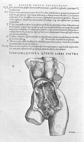
1 – [Anatomy of the Female Urogenital System]. From Vesalius, Andreas, De humani corporis fabrica libri septem (Basel: Joannem Oporinum, [1555]). Wellcome.
The early modern period saw an increased interest in anatomy that was expressed in illustrated anatomical books. A conviction began to grow among physicians that they should dissect and investigate the body themselves, rather than trusting to the wisdom of ancient authors. Andreas Vesalius made this argument in his book of 1543, De humani corporis fabrica, which was very widely translated, copied, and republished all over Europe. The woodcut illustrations in Vesalius’s book communicated new anatomical knowledge, but also made a rhetorical argument for the importance of direct observation through their newly detailed, more “naturalistic” style. [Read more]
Many anatomical illustrations from this period, including those made for Vesalius, were modeled on classical sculptures, which lent authority and visual context to the new knowledge. Others showed beautiful and apparently still living figures partly or completely anatomized, often exposing their own interiors. These images aimed to deflect the visceral horror associated with the dead dissected body. Such images played an important part in gathering and disseminating anatomical knowledge, but they were also used in many other ways by a variety of viewers who might see them primarily as erotic, as allegorical or moral statements, or simply as curiosities. [Read more]
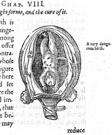
2 – [Birth Figure]. From Rüff, Jakob, The Expert Midwife, or an Excellent and Most Necessary Treatise of the Generation and Birth of Man (London: Printed by E. G. for S. B., [1637]). Wellcome.
Linked to anatomical illustrations were those found in works on surgery and midwifery. These showed not the ideal but the injured or sick body, the tools of the surgeon, and the practitioner enacting interventions upon the body: from removing a cataract, to bloodletting, to delivering a baby. For some people, these images formed part of their medical education. For others they provided a more abstract message about the specialist skills of the surgeon or midwife.
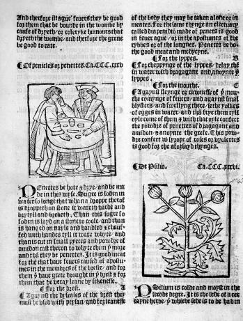
3 – [An Apothecary Administering Penettes] and [Psyllium]. From Anon., The Grete Herball Whiche Gyueth Parfyt Knowlege and Understandyng of all Maner of Herbes and there Gracyous Vertues ([London] : [P. Treveris for L. Andrewe?], [1529?]). Wellcome.
Botanical works were also often heavily illustrated, the images varying between highly observational and more symbolic, and between minutely detailed and very simple. Often color was added to prints by hand to make them more informative, as well as more beautiful and valuable. John Gerard’s Herball (1597) was a popular English herbal of the seventeenth century and contained many fine woodcut illustrations copied from earlier continental works. [Read more]
A system was developed in this time for representing all of a plant’s life-stages on a single specimen, including leaves, buds, flowers, and seeds together as never would be seen in nature. This system is still often used in botanical books today. It was disseminated in the early modern period through the widespread and accepted practice of copying and adapting existing images. While occasional battles over the stealing of images did flare up, more commonly the copying of an image was understood as a positive affirmation of its authority. Like anatomy books, herbals were used by different people for different purposes. A wealthy housewife, for instance, might use one as a reference work both for making remedies, and for designing embroidery patterns.
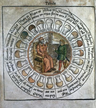
4 – Uroscopy chart. From Pinder, Ulrich, Epiphanie medicorum ([Place of publication not identified]: [publisher not identified], 1506). Wellcome.
Not all medical images were representations of things seen: they could be abstract charts or diagrams that visualized knowledge in other ways. One popular example is the uroscopy chart. Examining the color, consistency, and sediments in urine was a very common diagnostic practice in the early modern period. [Read more]
Uroscopy charts depicted multiple glass flasks of urine, each hand-colored and accompanied by text describing associated illnesses. These charts were first produced in medical manuscripts and while they were later transferred to printed works, they still required skilled hand-coloring. Astrological charts and diagrams were also very common. These helped with charting the position of stars and planets at particular times, and with drawing up horoscopes. For many early modern people, horoscopes were a serious business. Calculated to the specific moment of birth, they could say a lot about one’s bodily health and future fate. [Read more]
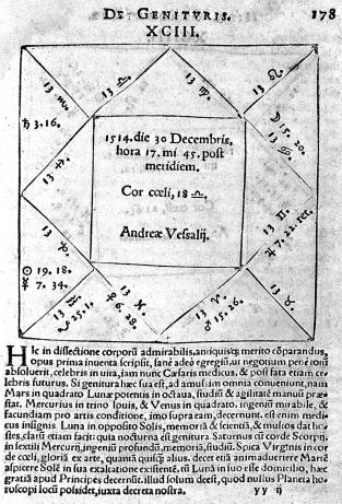
5 – [Horoscope of Andreas Vesalius]. From Cardano, Girolano, Libelli quinque (Nuremberg: J. Petreius, 1547). Wellcome.
Books with no other illustrations might contain a frontispiece—an image at the front of a book, often incorporating information such as the title and author’s name. These images communicated what kind of knowledge was in the book, but also argued for the legitimacy and importance of the author. Many represented the tools of the medical trade, anatomical figures, or portraits of the author or other famous physicians. They often adopted conventions from the fine arts, including allegorical figures, classical architectural elements, and fluttering angels. Such frontispieces say a lot about what the author wanted a reader to think about them and their work. [Read more]
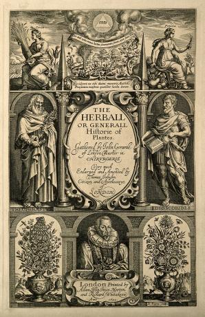
6 – Payne, J., Frontispiece. From Gerard, John, The Herball or, Generall Historie of Plantes (London: Adam Islip, Joice Norton and Richard Whitakers, 1633). Wellcome.
Images not only represented medical knowledge, they could also be medicines in themselves. Pregnant women were widely understood to be able to shape their unborn children through the power of “maternal imagination,” and so turned their gazes on images of healthy, hearty male children as models to replicate. [Read more]
Images could also be conduits for divine power: particularly (though not exclusively) in Catholic communities, printed images of saints were touched, kissed, and even swallowed in order to effect healing. [Read more]
Images are a rich historical resource, but they can also be tricky. They are not uncomplicated documentary evidence for what the past looked like, but have to be examined carefully for how they represent a complicated and (to us) strange culture. They can show what was most interesting, important, or contentious to the culture that produced them; what aspects of the medical profession were most valued; what representational styles and methods were in vogue; and how early modern people incorporated printed images into their everyday lives.
Further Reading
Dackerman, Susan, ed. Prints and the Pursuit of Knowledge in Early Modern Europe (New Haven and London: Yale University Press, 2011).
Fissell, Mary, “Hairy Women and Naked Truths: Gender and the Politics of Knowledge in ‘Aristotle’s Masterpiece’,” The William and Mary Quarterly, 60:1 (2003), 43-74.
Ivins, William. M. Jr., Prints and Visual Communication (Cambridge, MA and London: The MIT Press, 1978 [1953]).
Karr Schmidt, Suzanne, Altered and Adorned: Using Renaissance Prints in Daily Life (New Haven and London: Yale University Press, 2011).
Kusukawa, Sachiko, Picturing the Book of Nature: Image, Text, and Argument in Sixteenth-Century Human Anatomy and Medical Botany (Chicago and London: The University of Chicago Press, 2012).
Image Databases
Wellcome Collection Images https://wellcomecollection.org/works
Digitaler PortraitIndex http://www.portraitindex.de/
British Printed Images to 1700 http://www.bpi1700.org.uk/jsp/
US National Library of Medicine https://www.nlm.nih.gov/hmd/ihm/index.html
Anatomia Collection: Anatomical Plates 1522–1867 https://anatomia.library.utoronto.ca/
Historical Anatomies on the Web https://www.nlm.nih.gov/exhibition/historicalanatomies/intro.html
British Museum Collections Online https://www.britishmuseum.org/research/collection_online/search.aspx
Science Museum Collection http://collection.sciencemuseum.org.uk/